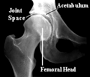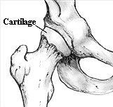

| An X-Ray and Illustration Showing a Normal Hip Joint |
Arthritis of the Hip Joint copyright ©1995 Herbert D. Huddleston, M.D.
For more information send email to moreinfo@scoi.com
| ANATOMY OF THE NORMAL HIP JOINT |
The hip joint is located where the thigh bone (femur) meets the pelvic bone. It is a ball and socket joint. The upper end of the femur is formed into a round ball (the head of the femur). A cavity in the pelvic bone forms the socket (acetabulum). The ball is normally held in the socket by very powerful ligaments that form a complete sleeve around the joint (the joint capsule). The capsule has a delicate lining (the synovium). The head of the femur is covered with a layer of smooth cartilage which is a fairly soft, white substance about 1/8 inch thick. The socket is also lined with cartilage (also about 1/8 inch thick). The cartilage cushions the joint, and allows the bones to move on each other with very little friction. An x-ray of the hip joint usually shows a space between the ball and the socket because the cartilage does not show up on x-rays. In the normal hip this joint space is approximately 1/4 inch wide and fairly even in outline.


| An X-Ray and Illustration Showing a Normal Hip Joint |