  |
Arthritis of the Knee Joint copyright ©1995 Herbert D. Huddleston, M.D.
For more information send email to moreinfo@scoi.com
Source:
![]()
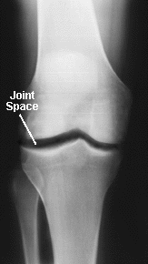
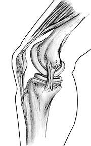
| ANATOMY OF THE NORMAL KNEE JOINT |
The knee is a "hinge type" joint which is formed by two bones held together by flexible ligaments. The bones are the femur (thigh bone) and the tibia (shin bone). The knee cap (patella) also forms part of the knee joint. It glides over the end of the femur as the knee bends. The moving parts of a normal knee are covered with a layer of articular cartilage which is a white smooth substance about 1/4 of an inch thick on the patella and 1/8 of an inch thick on the femur and tibia. An x-ray of the knee normally shows space (the "joint space") between the femur and the tibia as well as between the femur and the patella. This is not empty space but represents the cartilage (which does not show up on x-rays). The smooth, cartilage-covered surfaces of the knee move on each other with very little friction in the normal joint. In the normal knee the "joint space" is approximately 1/4 of an inch wide and fairly even in outline.
|
|
Source:
![]()