 Medial Side
 Medial Side
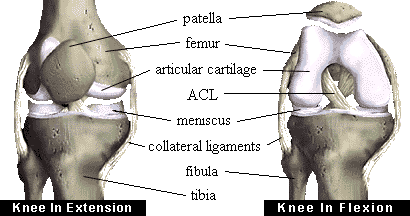
The bones of the knee, the femur and the tibia, meet to form a hinge joint. The joint is protected in front by the patella (kneecap). The joint is cushioned by articular cartilage that covers the ends of the tibia and femur, as well as the underside of the patella. The lateral meniscus and medial meniscus are pads of cartilage that further cushion the joint, acting as shock absorbers between the bones.
Ligaments help to stabilize the knee. The collateral ligaments run along the sides of the knee and limit sideways motion. The anterior cruciate ligament, or ACL, connects the tibia to the femur at the center of the knee. Its function is to limit rotation and forward motion of the tibia. (A damaged ACL is replaced in a procedure known as an ACL Reconstruction.) The posterior cruciate ligament, or PCL (located just behind the ACL) limits backward motion of the tibia.
These components of your knee, along with the muscles of your leg, work together to manage the stress your knee receives as you walk, run and jump.For more information on knee anatomy and knee disease.
| Other anatomy topics... | ||||||
|---|---|---|---|---|---|---|
| Hip | Shoulder | Ankle | Hand | Elbow | Spine | Great Toe! |